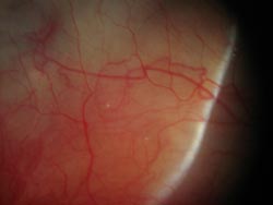
Scleritis
What is scleritis?
 Scleritis is inflammation of the tough, white structural wall of the eyeball, the sclera. The sclera is made of collagen and is continuous with the cornea, the clear window through which we see that makes up the front wall of the eye. Blood vessels run along and sometimes through the sclera, and can contribute to inflammation. The thin outside layer of the sclera, the episclera, can also be inflamed, but episcleritis is typically neither as severe nor symptomatic as scleritis. Swelling and severe inflammation of sclera can occur in one or both eyes, can affect surrounding tissues, and be quite dramatic and dangerous to vision.
Are there different types of scleritis?
There are several types of scleritis:
- Anterior
- Sectoral or Diffuse
- Nodular
- Necrotizing
- Posterior
Scleritis can be either anterior, in the front of the eye and visible during exam, or posterior, behind the eye and not visible during exam. Anterior scleritis can be sectoral or diffuse, depending on how much of the visible sclera is affected. It can also be nodular, presenting as a focal mound or elevation of inflamed tissue. Necrotizing scleritis, or scleromalacia perforans, is considered the most severe form of scleritis, and can cause dangerous thinning, potentially leading to perforation and loss of the eye. Even more alarming is the fact that necrotizing scleritis can at times present with voracious inflammation and be obvious and symptomatic, but at other times can be asymptomatic without obvious inflammation, with progression unknown to the patient until seen by the ophthalmologist. Scleritis can also occur in conjunction with inflammation of uvea, cornea, or other parts of the eye.
Causes of scleritis
Scleritis is most often idiopathic, or of unknown cause to the ophthalmologist, despite diagnostic measures. Autoimmune inflammation and infection are the two main causes, though trauma can be an inciting factor. Deposition of immune-complexes, or particles comprised of antibodies bound to another molecule (antigen), drive inflammation in a given area or sclera. The distinction between episcleritis and scleritis is of particular concern to the ophthalmologist – episcleritis is a benign condition where as scleritis can sometimes be a presenting sign of dangerous, and potentially fatal, underlying systemic disease.
Symptoms of scleritis
- Redness
- Pain
- Blurry vision
- Lid swelling
Redness may be isolated to a particular area of the eye, or diffuse. Pain can be excruciating and made worse with eye movement. Vision can be affected if swelling from inflammation affects surrounding tissues such as lens, cornea, choroid, retina, or optic nerve. Light sensitivity is not usually a symptom unless cornea (keratitis) is also involved.
What other medical conditions are associated with scleritis?
As stated above, scleritis can be a sign of more ominous systemic diseases. Non-infectious causes include:
- Rheumatoid Arthritis
- Systemic Lupus Erythematosus
- Inflammatory Bowel Disease •
- Relapsing Polychondritis
- Ankylosing Spondylitis
- Gout
- Reactive Psoriatic Arthritis
- Granulomatosis with Polyangiitis (formerly Wegener’s Granulomatosis)
- Microscopic Polyarteritis
- Churg-Strauss Syndrome
Infectious causes include:
- Herpes simplex
- Syphilis
- Bacteria
- Tuberculosis
- Fungi
How is scleritis diagnosed?
As with all ocular inflammation, a careful history and review of systems is done. Note is taken of potential systemic issues which have symptoms revealed on review of systems, i.e. joint swelling, rash, abdominal pain. Examination reveals inflammation of deep scleral vessels, or areas of necrosis (cell death) and scleral thinning, which can be photographed for the record. Eye drops may be able to more easily distinguish between inflammation of sclera and episclera when it is unclear. Posterior inflammation is usually not visible on exam, and the ophthalmologist can use ultrasound, looking for signs of inflammation behind the eye. Occasionally changes in retina or optic nerve appear from posterior inflammation. Serologic evaluation is typically performed to search for possible autoimmune or infectious causes. Lastly, scleral biopsy with microscopic evaluation of prepared tissue can give important information on specific patterns of inflammation seen and the presence or absence of certain infectious organisms.
What are complications from scleritis?
The most dreaded complication of scleritis is perforation, which can lead to dramatic vision loss, infection, and loss of the eye. Damage to other inflamed areas, such as cornea or retina, may leave permanent scarring and cause blurring. Chronic pain can be debilitating if not treated. Patients may suffer complications from treatment more often than disease itself, with development of cataract or secondary glaucoma from chronic corticosteroid use.
How do you treat scleritis?
Treatment should be aimed at quieting inflammation quickly. Antibiotic therapy can be used when an infectious cause is shown or even highly suspected, along with topical corticosteroids for some infections (never fungus). For non-infectious causes, oral or topical corticosteroids can be used, as well as oral or topical non-steroidal anti-inflammatory drugs (NSAIDs). Periocular corticosteroid injection is a debated subject, as some fear that areas of necrosis are at higher risk for melt and perforation, though evidence suggests that treatment of non-necrotizing scleritis with injections is effective. Intravenous steroids can also be used. Of course, dependence on steroid therapy should be avoided due to complications of long term steroid use.
Systemic therapy, by mouth, injection, or intravenous infusion, is necessary to treat chronic or recurrent scleritis. Immunomodulatory therapy, utilizing a step-ladder approach, can be used effectively for non-infectious scleritis, with regular monitoring for blood work and side effects. Surgery to repair perforated sclera, or bolster dangerously thinned sclera, can be done with prepared scleral grafts, or other similar available sterile tissue.
Our Physicians
All of our physicians have completed Fellowships in their specialty.
C. Stephen Foster, MD, FACS, FACR
Founder
Stephen D. Anesi, MD, FACS
Partner and Co-President
Peter Y. Chang, MD, FACS
Partner and Co-President
Peter L. Lou, MD
Associate
