
What Does Retina Imaging Show? Understanding OCT and Other Tests
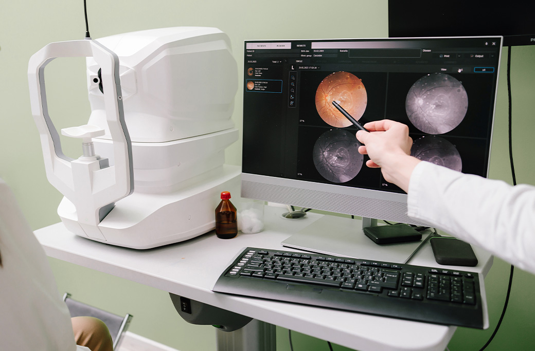
Retina imaging gives an eye doctor detailed information about the structures, layers, blood vessels and (in certain tests) function of the retina. Depending on the test, imaging may reveal retinal swelling, fluid, thinning, abnormal blood vessels, bleeding, inflammation, scar tissue or other changes that may not be fully visible during a routine eye examination.
No single test answers every question. A retina specialist selects imaging based on your symptoms, examination findings, medical history and the condition being evaluated.
Why Is Retinal Imaging Performed?
The retina is a thin layer of light-sensitive tissue lining the back of the eye. Its central area, called the macula, supports the detailed vision used for reading, driving and recognizing faces.
Since retinal conditions can affect different structures, a specialist may need more information than visual-acuity testing or a surface examination can provide. Retinal imaging can help the doctor:
-
-
Examine the layers of the retina and macula
-
Identify swelling or fluid in or beneath the retina
-
Evaluate retinal and choroidal blood vessels
-
Document changes for comparison at later visits
-
Investigate unexplained distortion or vision loss
-
Monitor a known condition
-
Assess whether a treatment is producing the intended anatomical response
-
Decide whether additional testing is needed
-
Imaging findings must be interpreted together with your symptoms, medical history and dilated examination. An image by itself does not always establish a diagnosis.
Learn more about retina care, diagnosis and treatment at MERSI.
What Does An OCT Scan Show?
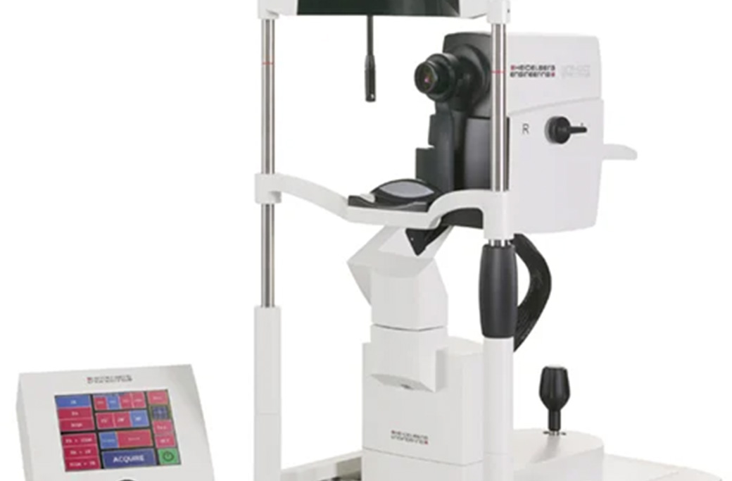
Optical coherence tomography, usually called OCT, uses light to create high-resolution, cross-sectional images of the retina. The resulting images allow the doctor to examine individual retinal layers in a way that is sometimes compared with viewing a side profile or a layered map of the tissue.
An OCT scan may help show:
-
-
Retinal or macular swelling
-
Fluid within or beneath the retina
-
Changes in retinal thickness
-
Separation between retinal layers
-
An epiretinal membrane or macular pucker
-
A macular hole
-
Changes associated with macular degeneration
-
Changes related to diabetic retinal disease
-
Structural effects of retinal inflammation
-
Certain abnormalities involving the optic nerve or retinal nerve-fiber layer
-
What a particular OCT finding means depends on its location, pattern and clinical context. Similar symptoms can have different causes, and different retinal conditions can sometimes produce similar-looking changes.
Does an OCT scan use radiation?
OCT uses light rather than X-rays. It does not expose the eye to ionizing radiation.
Is an OCT scan painful?
OCT is noninvasive. During the scan, you generally place your chin and forehead against supports and look at a target while the instrument captures images. Nothing is injected into the eye for a standard OCT scan.
The technician may repeat a scan if blinking or eye movement affects image quality. Whether your pupils need to be dilated depends on the examination, the instrument and the images your doctor needs.
What Is The Difference Between OCT And A Retinal Photograph?
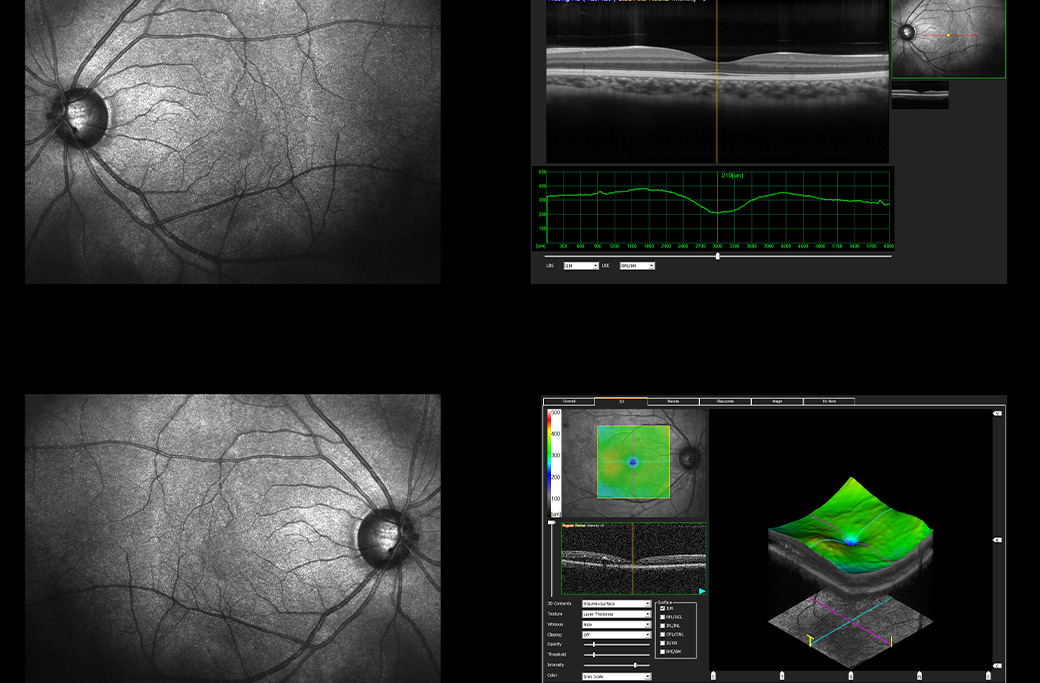
An OCT scan and a retinal photograph show different views of the eye.
A retinal or fundus photograph records the visible appearance of the back of the eye. It can document features such as:
-
-
The optic nerve
-
The macula
-
Retinal blood vessels
-
Pigment changes
-
Bleeding or deposits
-
Certain lesions or areas of retinal damage
-
These photographs create a record that can be compared with images from later visits.
OCT provides cross-sectional information about retinal layers and thickness. A retinal photograph shows the surface appearance of a wider area. The tests can therefore complement each other rather than serve as interchangeable alternatives.
What Does Fluorescein Angiography Show?
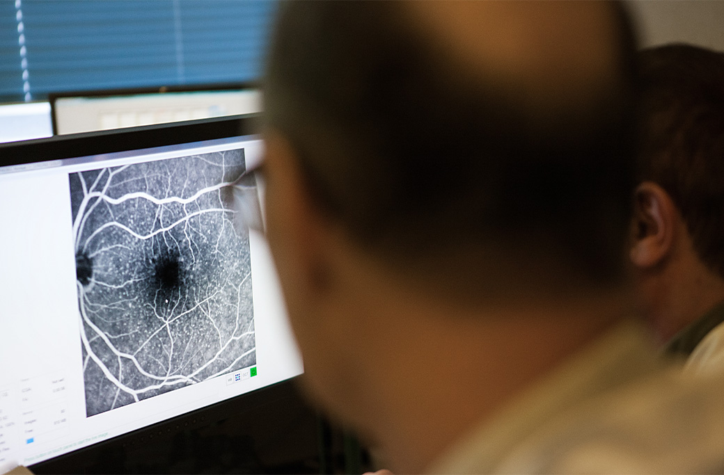
Fluorescein angiography helps the doctor evaluate blood flow through retinal vessels. A fluorescent dye is injected into a vein, usually in the arm or hand. As the dye circulates through the blood vessels in the eye, a specialized camera takes a sequence of photographs.
The test may help identify:
-
-
Leaking retinal blood vessels
-
Areas of blocked or reduced circulation
-
Abnormal blood-vessel growth
-
Vascular changes related to diabetic retinopathy
-
Findings associated with retinal vein occlusion
-
Patterns of leakage or inflammation
-
Blood-vessel changes associated with certain macular conditions
-
Fluorescein angiography provides information about circulation and leakage that a standard OCT scan may not show in the same way.
Since this test involves an injected dye, tell the clinical team about your medical history, previous reactions to injected dyes or medicines, and any other information they request before the procedure. The team should explain what you may experience and any applicable precautions.
What Is ICG Angiography?
Indocyanine green angiography, or ICG angiography, is another dye-based test. It is used to examine circulation in the choroid, the blood-vessel-rich layer beneath the retina.
A specialist may recommend ICG angiography when the clinical question involves deeper choroidal circulation or when other imaging has not provided enough information.
Fluorescein and ICG angiography are not automatically required for every patient. The specialist determines whether either test is appropriate based on the condition being investigated and the information already available.
What Is Fundus Autofluorescence?
Fundus autofluorescence uses naturally occurring fluorescence within retinal tissue to create an image. It can help document patterns involving the retinal pigment epithelium, a layer of cells that supports the retina.
Depending on the condition, autofluorescence may help the doctor evaluate or monitor areas where these supporting cells appear stressed, altered or damaged.
The colors or bright and dark areas in an autofluorescence image are not a direct picture of how a patient sees. They are patterns that require clinical interpretation.
What Can Eye Ultrasound Reveal?
Ocular ultrasound uses sound waves to create images of structures inside the eye. MERSI’s public testing information includes A-scan and B-scan ultrasound.
B-scan ultrasound may be useful when the doctor cannot obtain a clear view through the front of the eye or when additional structural information is needed. Depending on the clinical situation, it may help evaluate the retina, vitreous and other internal structures.
Ultrasound does not replace every other retinal test. It answers different questions and may be combined with examination findings or other imaging.
What Do Microperimetry And Electrophysiological Tests Measure?
Most retinal imaging tests primarily examine structure. Other tests can provide information about visual function.
Microperimetry
Microperimetry assesses visual sensitivity in specific retinal areas, including the fovea. It can help the doctor compare a patient’s visual response with a particular retinal location.
Electrophysiological Testing
Electrophysiological testing evaluates how parts of the visual system respond to light or visual stimulation. Depending on the test, it may provide information about retinal function or the visual pathway from the retina toward the brain.
These tests are not routine for every retina patient. They may be considered when symptoms, examination findings and structural images do not provide a complete explanation.
MERSI’s advanced eye testing and diagnostic services page provides an overview of testing currently described by the practice.
Can Retinal Imaging Detect A Problem Before Symptoms Begin?
Imaging can sometimes document structural or vascular changes before a patient notices a meaningful change in vision. That does not mean every retinal condition can be detected by imaging alone or that imaging replaces a comprehensive eye examination.
Some retinal diseases cause few noticeable early symptoms. The appropriate combination of examination, dilation and imaging depends on a person’s diagnosis and risk factors.
Patients with diabetes, macular degeneration, retinal vascular disease, ocular inflammation or another known retinal condition may require repeated imaging so their doctor can compare findings over time.
Can Imaging Determine Whether Treatment Is Working?
Retinal imaging can provide objective information about anatomical changes during treatment or monitoring. For example, a specialist may compare images to evaluate whether swelling or fluid has changed.
An improved scan does not always mean that vision will improve by the same amount. Likewise, a person’s vision may feel stable even when imaging shows a change that deserves attention.
Treatment decisions should therefore be based on the complete clinical picture rather than one measurement or image.
Will Every Retina Appointment Include The Same Tests?
No. Testing is individualized.
One patient may need a dilated examination and OCT, while another may also need retinal photography, angiography, ultrasound or functional testing. The choice can depend on:
-
-
The symptoms being investigated
-
The suspected or confirmed diagnosis
-
Whether the condition is new or already being monitored
-
Whether the doctor needs to examine structure, circulation, function or a combination
-
The quality of earlier images
-
Whether the result could affect monitoring or treatment decisions
-
If you are unsure why a test was ordered, ask what clinical question it is intended to answer and how the result may affect your care.
How Should You Prepare For Retinal Imaging?
Preparation depends on the test.
Before your visit:
-
-
Bring an up-to-date medication list.
-
Tell the team about relevant medical conditions and prior reactions to medicines or injected dyes.
-
Bring glasses or other vision aids you normally use.
-
Ask whether your pupils may be dilated.
-
Consider arranging transportation if dilation or your current symptoms could make driving difficult.
-
Follow any test-specific instructions supplied by the practice.
-
Pupil dilation can temporarily cause light sensitivity and blurred near vision. The duration and effect vary, so follow the clinical team’s advice about driving and other activities after the visit.
What Questions Should You Ask About Your Results?
Useful questions include:
-
-
Which retinal structures did this test examine?
-
Did the scan show fluid, swelling, thinning or another change?
-
Is the finding new, stable or progressing?
-
Does the image explain my symptoms?
-
Do I need another type of imaging?
-
How often should this test be repeated?
-
Will the result change my treatment or monitoring plan?
-
Which symptoms should prompt an earlier call?
-
The specialist should interpret the images in the context of your examination and explain what the findings mean for your individual care.
Schedule A Retina Evaluation At MERSI
MERSI provides retinal evaluation and diagnostic testing in Waltham for patients from Greater Boston and throughout New England.
If you have a known retinal condition, a referral for retinal imaging or an unexplained change in vision, you may request an appointment with MERSI or call 781-891-6377.
Sudden vision loss, new flashes with a rapid increase in floaters, or a curtain or shadow in the visual field may require urgent evaluation. Do not rely on a routine online appointment request for a possible vision emergency.
This article provides general educational information and does not replace an examination, diagnosis or individualized medical advice from a qualified healthcare professional.
Frequently Asked Questions
What diseases can an OCT scan detect?
OCT can document structural changes associated with several retinal and optic-nerve conditions, including macular degeneration, diabetic macular edema, epiretinal membranes, macular holes and certain inflammatory conditions. An OCT image does not diagnose every condition by itself; the doctor must interpret it with the examination and medical history.
Is OCT the same as a retinal photograph?
No. OCT creates cross-sectional images of retinal layers. A retinal photograph documents the visible appearance of the back of the eye. The tests provide different, often complementary information.
Can OCT show a retinal tear or detachment?
OCT may show certain separations, fluid or structural changes in the area scanned, particularly near the macula. Evaluating a suspected peripheral retinal tear or detachment generally also requires a careful dilated examination and, in some circumstances, additional imaging such as ultrasound. A normal central OCT should not be used by a patient to rule out a peripheral retinal problem.
Why would I need fluorescein angiography if I already had OCT?
OCT primarily provides structural information. Fluorescein angiography provides information about retinal circulation, leakage and blocked or abnormal blood vessels. A specialist may use both when the tests answer different clinical questions.
How long does retinal imaging take?
The timing varies with the test, the number of images required, whether dilation is needed and whether injected dye is used. The practice can provide more specific scheduling and preparation information when your test is ordered.
Medically reviewed by:
Stephen D. Anesi, MD, FACS, Partner & Co-President
Specializes in: Ocular inflammatory diseases, cornea, glaucoma (medical, surgical & MIGS), cataract, and medical retina
Peter Y. Chang, MD, FACS, Partner & Co-President
Specializes in: Ocular inflammatory diseases, vitreoretinal surgery, and cataract
Carl S. Wilkins, MD
Specializes in: Ocular inflammatory diseases, vitreoretinal surgery, and cataract
Reviewed: August 27, 2026