
Uveitis is a complex inflammatory eye condition that requires more than a basic vision test to diagnose. Since it can be linked to underlying autoimmune diseases or infections, your eye doctor may recommend a series of specialized tests to understand both the location and cause of inflammation.
Early and accurate diagnosis is essential to prevent vision loss and begin the right treatment as quickly as possible. To better understand the symptoms, causes, and treatment options associated with inflammatory eye disease, read our guide on What Is Uveitis? Symptoms, Causes & When to See a Specialist.
Why Are So Many Tests Needed for Uveitis?
Uveitis is not a single disease. It is a sign of inflammation inside the eye that can have multiple causes. In some patients, it is related to autoimmune conditions, while in others it may be triggered by infection or trauma.
Due to this complexity, testing serves two key purposes:
-
- Confirm the presence of inflammation inside the eye
- Identify the underlying cause of that inflammation
A targeted diagnosis helps guide the most effective treatment plan. Several advanced diagnostic tools may be used during your evaluation. Learn more about the specific imaging and laboratory studies in How Is Uveitis is Diagnosed: Tests and Eye Exam Process.
What Happens During a Comprehensive Eye Examination?
The first step in diagnosing uveitis is a complete eye exam performed by an ophthalmologist or retina specialist.
This typically includes:
-
- Visual acuity testing (checking how well you see)
- Eye pressure measurement
- Evaluation of eye redness and irritation
- Detailed medical history review
While this step does not confirm uveitis on its own, it helps guide further testing.
What Does a Slit-Lamp Examination Show?
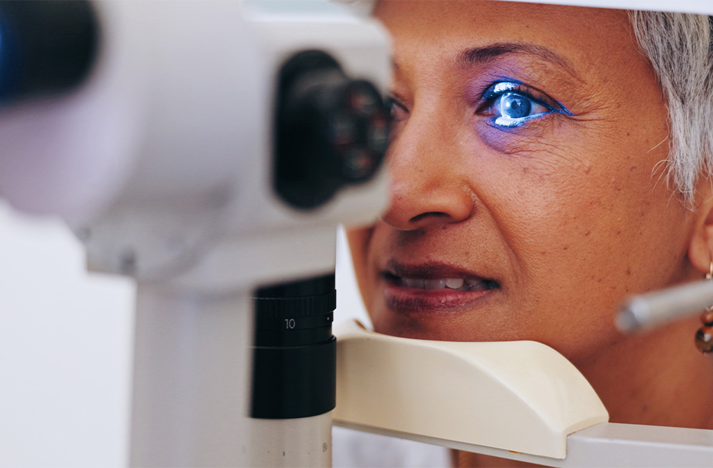
A slit-lamp exam is one of the most important tools in diagnosing uveitis.
This microscope allows your doctor to examine the front structures of the eye in detail. It can detect:
-
- Inflammatory cells in the anterior chamber
- Protein buildup caused by inflammation
- Signs of anterior uveitis
This test is painless and performed during a routine office visit.
Dilated Eye Exam
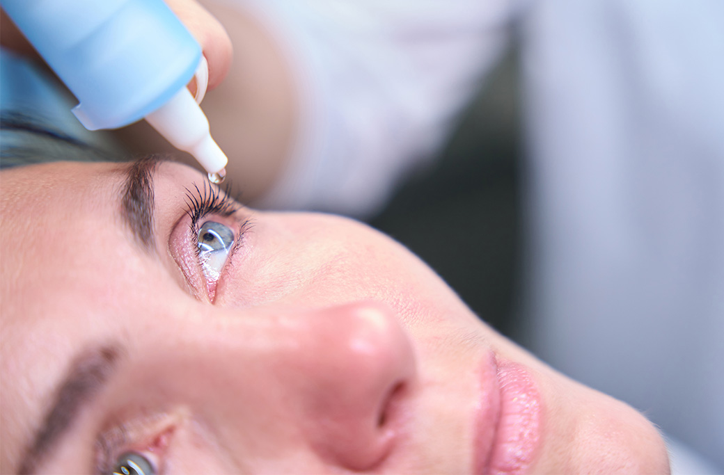
A dilated exam allows your specialist to examine the back of the eye, including the retina and optic nerve.
This test helps identify:
-
- Posterior uveitis
- Retinal swelling (macular edema)
- Optic nerve inflammation
- Vascular changes in the retina
Dilating drops are used to temporarily widen the pupil for a better view of internal eye structures.
How Does OCT Help Diagnose Uveitis?
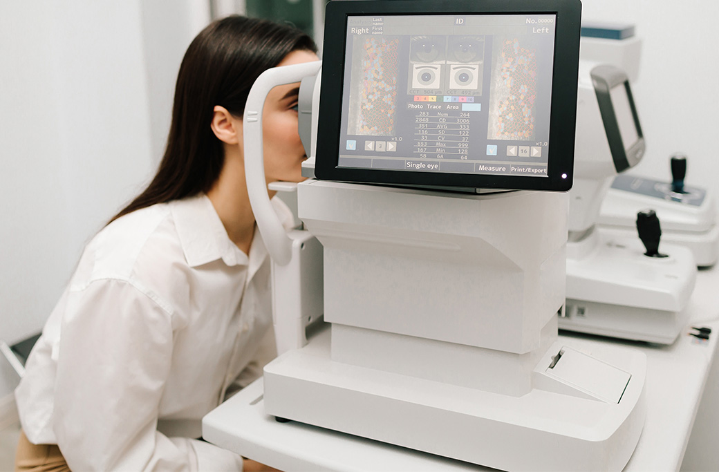
OCT is a high-resolution imaging test that provides cross-sectional images of the retina.
It is commonly used to detect:
-
- Macular edema (retinal swelling)
- Fluid buildup in the retina
- Structural changes caused by inflammation
OCT is quick, non-invasive, and one of the most important tools for monitoring disease progression.
Why Is Fluorescein Angiography Used for Uveitis?
Fluorescein angiography is used to evaluate blood flow in the retina.
During the test:
-
- A fluorescent dye is injected into the bloodstream
- A specialized camera captures images as the dye travels through retinal blood vessels
This helps detect:
-
- Blood vessel leakage
- Retinal inflammation
- Areas of poor circulation
Blood Tests and Laboratory Work
Since uveitis is often associated with systemic disease, blood work may be needed to identify underlying conditions.
Common tests include:
-
- Autoimmune markers (such as ANA, RF, HLA-B27)
- Inflammatory markers
- Infectious disease testing (Lyme disease, tuberculosis, syphilis)
- General health screening panels
These tests help determine whether the inflammation is related to an autoimmune disorder or infection elsewhere in the body.
Imaging Beyond the Eye
In some cases, additional imaging may be recommended if a systemic condition is suspected.
This may include:
-
- Chest X-rays (to evaluate for sarcoidosis or tuberculosis)
- MRI or CT scans in specific cases
- Rheumatologic evaluation
Since uveitis can be linked to diseases affecting the entire body, collaboration with other specialists is sometimes necessary.
How Long Does Uveitis Testing Take?
Some test results are available immediately during your visit, such as slit-lamp or OCT imaging. However, blood tests and systemic evaluations may take several days to return.
In more complex cases, your doctor may continue ordering tests over time to better understand the underlying cause.
When Should You Be Tested for Uveitis?
You should seek evaluation if you experience:
-
- Persistent eye redness
- Eye pain or pressure
- Sensitivity to light
- Blurred vision
- New floaters or vision changes
Since uveitis can progress quickly, early testing is essential for protecting long-term vision.
Uveitis Testing at MERSI
At MERSI, uveitis evaluation is supported by advanced diagnostic technology and specialized expertise in ocular inflammatory disease.
Our team uses a combination of imaging, clinical examination, and laboratory testing to identify both the presence and cause of inflammation.
This comprehensive approach allows for faster diagnosis and more precise treatment planning.
Key Takeaway
There is no single test for uveitis. Diagnosis requires a combination of eye exams, imaging, and laboratory testing to confirm inflammation and identify its cause. Early evaluation is essential to prevent vision loss and begin appropriate treatment.
When to Seek Care
If you are experiencing eye pain, redness, light sensitivity, or sudden vision changes, you should be evaluated by a uveitis specialist as soon as possible.
Schedule an Evaluation at MERSI
At MERSI, our retina and uveitis specialists use advanced imaging and diagnostic testing to quickly identify the cause of eye inflammation and create a personalized treatment plan.
If you are concerned about uveitis or unexplained eye symptoms, schedule a consultation with our team in Waltham, MA.
Frequently Asked Questions
What tests are used to diagnose uveitis?
Uveitis is diagnosed using a combination of eye exams and specialized imaging tests, including slit-lamp examination, dilated eye exam, OCT imaging, fluorescein angiography, and blood work to identify underlying causes.
Can uveitis be diagnosed with a regular eye exam?
A routine eye exam may detect signs of inflammation, but a full uveitis diagnosis requires specialized testing such as slit-lamp evaluation, retinal imaging, and sometimes laboratory testing.
What is the most important test for uveitis?
The slit-lamp examination is one of the most important tests because it allows doctors to directly see inflammatory cells inside the eye.
Why do doctors order blood tests for uveitis?
Blood tests help identify underlying conditions such as autoimmune diseases or infections that may be causing eye inflammation.
Does diagnosing uveitis require imaging tests?
Yes. Imaging tests like OCT and fluorescein angiography help detect retinal swelling, inflammation, and blood vessel leakage that may not be visible during a standard exam.
How long does it take to diagnose uveitis?
Some findings are immediate during an eye exam, but full diagnostic testing (including blood work) may take several days depending on the complexity of the case.