
How Is Uveitis Diagnosed? Tests & Eye Exam Process Explained
Quick answer: Uveitis is diagnosed through a comprehensive eye examination combined with a systemic medical evaluation. Ophthalmologists use slit-lamp exams, dilated fundus evaluations, and advanced imaging like optical coherence tomography (OCT) to locate inflammation. Blood work and laboratory tests are then used to identify underlying autoimmune or infectious causes.
When eye inflammation threatens your vision, pinpointing the exact cause requires more than a standard vision check. Uveitis requires a comprehensive eye exam combined with a thorough systemic evaluation. The diagnostic process at the Massachusetts Eye Research and Surgery Institution (MERSI) goes beyond visual inspections to include high-resolution imaging and detailed laboratory work. Since uveitis can progress rapidly and cause permanent damage, an early and accurate diagnosis is vital for preventing severe complications like vision loss. To understand the foundational risks of this condition, you can review our guide on What Is Uveitis? Symptoms, Causes & When to See a Specialist.
What Happens During an Initial Uveitis Clinical Evaluation?
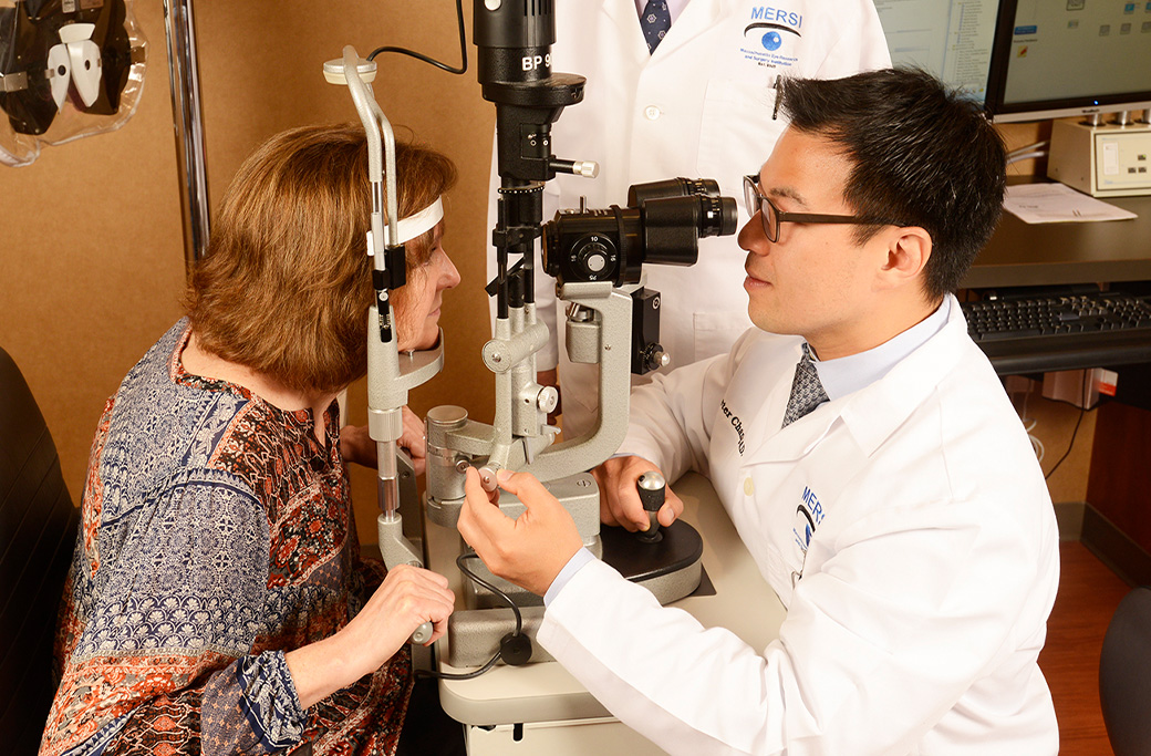
The diagnostic workflow begins the moment you enter the clinic. Ophthalmologists start by conducting a rigorous medical history review. Your doctor will ask about your current health, past medical conditions, and any family history of autoimmune diseases. This step provides vital clues about systemic conditions that might be driving the eye inflammation.
Next, the physician will guide a detailed symptom discussion. You will need to describe when your symptoms started, whether they affect one or both eyes, and the severity of your discomfort. Finally, the clinical team performs visual acuity testing to establish a baseline for your current eyesight. By measuring exactly how much your vision has been affected, the eye specialist can track your response to future treatments.
How Does A Slit-Lamp Examination Detect Anterior Uveitis?
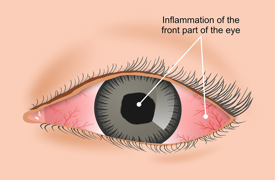
A slit-lamp examination serves as the core diagnostic tool for identifying inflammation in the front portion of the eye. This device is essentially a specialized microscope equipped with a bright, focused beam of light. During the exam, the ophthalmologist looks directly into the anterior chamber, which is the fluid-filled space between your cornea and iris.
The primary goal of this non-invasive test is to detect inflammatory white blood cells floating in the eye fluid. The presence and quantity of these cells confirm a diagnosis of anterior uveitis and help the doctor grade the severity of the flare-up. The slit-lamp exam also reveals protein flare and inflammatory debris, giving the specialist immediate visual evidence of the disease’s activity without requiring any surgical procedures.
What Does A Dilated Eye Exam Reveal About Posterior Uveitis?
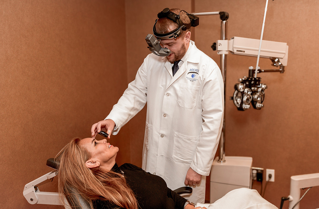
To evaluate the deeper structures of the eye, your ophthalmologist must perform a dilated eye exam and fundus evaluation. By applying special dilating drops, the doctor widens your pupil, creating a clear window to inspect the retina, the choroid, and the optic nerve.
This step is essential for identifying posterior uveitis and panuveitis. During the fundus evaluation, the specialist looks for specific signs of internal damage, such as retinal swelling, active lesions, or vascular changes. Since posterior uveitis carries a higher risk of permanent vision loss, this detailed inspection allows the physician to detect sight-threatening complications before they cause irreversible harm.
Which Imaging Tests Are Used To Diagnose Uveitis?
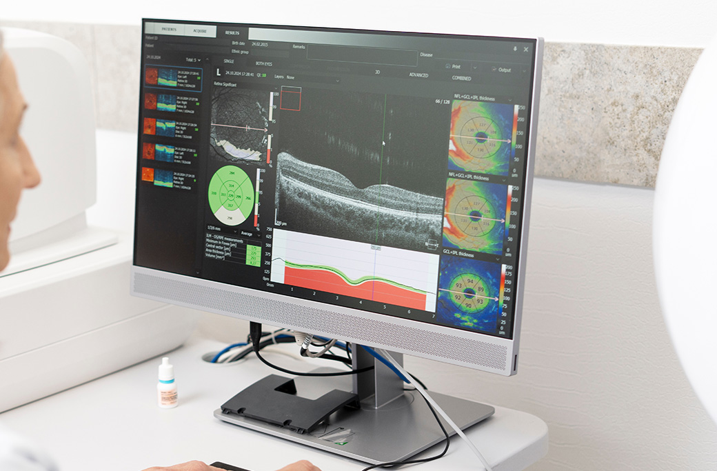
While physical examinations reveal surface-level and internal inflammation, advanced imaging tests provide a microscopic view of the eye’s structural health. These tools allow doctors to see precisely how inflammation is impacting cellular layers. For a deeper dive into the specific testing rationale, read our resource on What Tests Are Used to Determine the Cause of Uveitis?.
Optical Coherence Tomography (OCT)
Optical Coherence Tomography (OCT) is a high-resolution retinal imaging test that captures cross-sectional pictures of the retina. The OCT scan measures retinal thickness, making it highly effective at detecting macular edema (swelling in the central retina). Macular edema is a common and dangerous complication of uveitis, and OCT allows specialists to measure this swelling down to the micron.
Fluorescein Angiography
Fluorescein angiography involves injecting a fluorescent dye into a vein in your arm. As the dye travels through the blood vessels in your retina and choroid, a specialized camera takes rapid photographs. This test highlights blood vessel leakage and identifies areas of severe retinal inflammation. Choose fluorescein angiography if identifying precise areas of vascular damage matters more to the treatment plan than simply measuring overall tissue thickness.
What Laboratory Tests And Blood Work Check For Systemic Causes?
Due to uveitis frequently linking to diseases affecting the entire body, a comprehensive systemic workup is a mandatory phase of the diagnostic process. Laboratory testing and targeted blood work help the specialist uncover the root trigger of the eye inflammation.
Your ophthalmologist will typically order screening panels that check for autoimmune markers, infectious diseases, and general inflammatory markers. Common autoimmune conditions tested during this phase include Rheumatoid arthritis, Lupus, and Sarcoidosis. Additionally, the ophthalmologist will screen for infectious diseases like Lyme disease and Tuberculosis (TB). Identifying a positive marker in your blood work allows the medical team to treat the underlying systemic disease, rather than just temporarily suppressing the eye inflammation.
Why Do Eye Doctors Look For Underlying Causes Of Uveitis?
Uveitis is often a symptom of a larger health issue, rather than a standalone eye disease. The ultimate goal of a thorough diagnostic workup is identifying systemic conditions that might be attacking the patient’s immune system.
Finding an underlying cause completely changes the treatment strategy. If a patient has an infectious disease like Lyme disease, they require targeted antibiotics. If the patient has an autoimmune condition like Rheumatoid arthritis, they need immunosuppressive therapies. Understanding this root cause ensures the patient receives the correct interventions. You can explore these medical pathways in our overview of Uveitis Treatment Options or learn about advanced biologic therapies by reading What Does IV Treatment for Uveitis Feel Like?.
How Long Does A Complete Uveitis Diagnosis Take?
The timeline for reaching a definitive uveitis diagnosis varies based on the complexity of the patient’s condition. In straightforward cases, the ophthalmologist can provide a same-day diagnosis using the slit-lamp exam and OCT imaging. You leave the clinic with a clear understanding of the inflammation’s location and severity.
However, uncovering the exact systemic cause often requires laboratory results that take several days to process. Highly complex cases need multidisciplinary coordination between the eye specialist, rheumatologists, and infectious disease experts. This collaborative review can extend the diagnostic timeline to a few weeks.
When Should You Seek Immediate Medical Evaluation For Eye Inflammation?
Uveitis can cause permanent damage quickly, so recognizing the early warning signs is vital for your long-term visual health. You should seek an immediate evaluation from a specialist if you experience deep, aching eye pain that does not resolve.
Sudden light sensitivity, a rapid increase in floaters, or dark spots appearing in your visual field require urgent attention. Sudden vision changes, including profound blurring or a noticeable drop in visual acuity, indicate that internal eye structures are under active attack. Do not wait for these symptoms to clear up independently. Schedule a medical evaluation right away.
Why Choose MERSI For Your Uveitis Evaluation?
Securing an accurate uveitis diagnosis involves a complex but essential series of steps. Early evaluation and intervention are your best defenses to protect your vision from permanent damage. The Massachusetts Eye Research and Surgery Institution (MERSI) provides advanced diagnostic tools, in-house imaging, and a CLIA-certified pathology lab, allowing our specialists to evaluate your condition rapidly and thoroughly.
Take action today to safeguard your eyesight. Schedule a comprehensive evaluation and contact the Top Boston Retina & Uveitis Specialists | MERSI Waltham to get started.
Frequently Asked Questions about Uveitis Diagnosis
What is the most accurate test for diagnosing anterior uveitis?
The slit-lamp examination is the most accurate and commonly used test for diagnosing anterior uveitis. It allows the ophthalmologist to look directly into the anterior chamber of the eye under high magnification to detect white blood cells and protein flare.
Will I need a blood test to diagnose eye inflammation?
Yes, patients diagnosed with uveitis usually require blood tests. Because uveitis is frequently linked to systemic autoimmune diseases or infections, blood work helps doctors identify conditions like Lupus, Sarcoidosis, or Lyme disease that may be causing the eye inflammation.
Does a uveitis eye exam hurt?
The physical eye exams and imaging tests used to diagnose uveitis are generally painless. You will receive dilating drops which may cause temporary light sensitivity, and fluorescein angiography involves a standard needle stick in the arm to deliver the dye. The eye evaluations themselves remain non-invasive.