Cataracts vs Retinal Disease: What’s Causing Vision Changes?
Quick answer: Cataracts and retinal disease can both cause blurry vision, but the symptoms often differ in important ways. Cataracts typically cause gradual cloudy vision, glare, halos around lights, faded colors, and difficulty seeing at night. Retinal diseases are more likely to cause distorted vision, floaters, dark spots, missing areas of sight, or sudden vision changes. Since symptoms can overlap, a comprehensive eye evaluation is the best way to determine the true cause of vision problems.
When vision becomes blurry, many people assume cataracts are to blame, and often, they are. Cataracts are one of the most common age-related eye conditions and a leading cause of gradual vision changes. However, blurry vision does not always begin in the lens of the eye.
Sometimes, the issue lies in the retina (the light-sensitive tissue at the back of the eye responsible for processing what you see). Conditions such as macular degeneration, diabetic retinopathy, retinal swelling, and retinal tears can sometimes mimic cataract symptoms or develop quietly alongside them. In some cases, patients may even have both cataracts and retinal disease at the same time.
Understanding the difference between cataracts vs retinal disease can help you recognize when symptoms deserve medical attention and why identifying the true cause matters before treatment decisions (including cataract surgery) are made.
How Do Cataracts and Retinal Disease Affect Vision Differently?
To understand why symptoms sometimes look alike, it helps to know what each condition actually affects.
What cataracts do to your vision
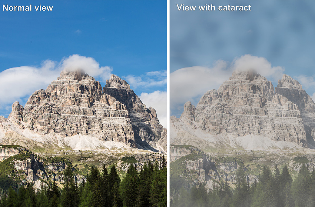
A cataract is a change in the clarity of the eye’s natural lens, the structure that focuses incoming light onto the retina. When the lens becomes clouded, light scatters before it ever reaches the back of the eye. The result is hazy, dimmed, or blurred vision that worsens over time.
What retinal disease does to your vision
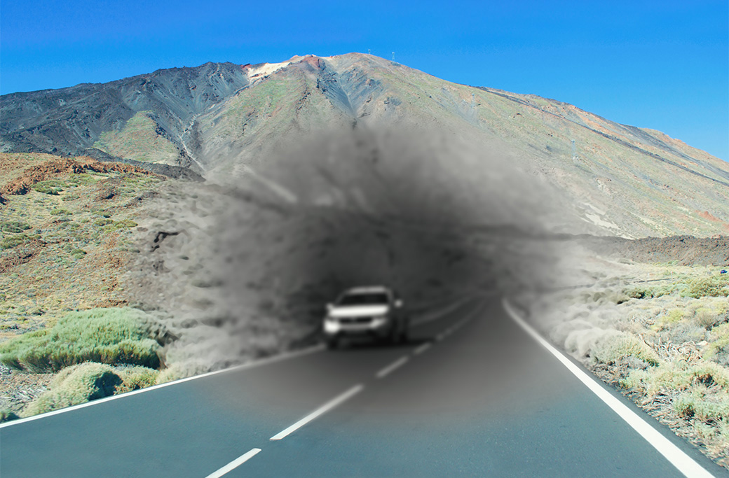
The retina doesn’t focus light, it processes it. This tissue contains millions of photoreceptor cells that convert light into electrical signals, which travel through the optic nerve to the brain. When the retina is damaged, the brain receives a distorted or incomplete image, regardless of how clearly the lens focuses incoming light.
Why the symptoms can overlap
Both cataracts and retinal disease can reduce overall visual clarity and make reading more difficult. The key difference is in the quality of the blur. Cataract-related blur tends to feel like looking through frosted glass—uniformly hazy. Retinal-related blur is more likely to involve distortion, gaps, or changes that feel sudden or uneven.
What Are the Most Common Symptoms of Cataracts?
Gradual, cloudy, or blurry vision
Cataract symptoms develop slowly, often over months or years. Patients frequently describe the experience as looking through a foggy or dirty window. The progression is painless, and many people don’t notice the change until vision has declined significantly.
Glare and halos around lights
As the lens becomes more opaque, light scatters in ways it shouldn’t. Bright lights, particularly headlights while driving at night or direct sunlight, often produce uncomfortable glare or visible halos.
Difficulty driving at night
Reduced contrast sensitivity is a hallmark cataract symptom. Low-light environments that require the eye to work harder become increasingly challenging, and many patients report losing confidence driving after dark before they ever seek an evaluation.
Faded or yellowed colors
The proteins that cloud the lens can also shift its color. Colors may appear less vibrant than they once did, and whites may take on a yellow or brownish tint.
Frequent changes in glasses prescription
In the early stages, cataract-related vision changes may be corrected with updated glasses. Over time, however, no prescription adjustment will fully compensate for the lens opacity. If new glasses are no longer helping the way they used to, it may be time to explore what’s happening inside the eye.
What Are the Most Common Symptoms of Retinal Disease?
This is where cataracts vs. retinal disease becomes especially important to understand, because retinal symptoms are often misread as something less urgent.
Distorted or wavy vision
One of the clearest signs of a retinal problem (particularly macular degeneration or retinal swelling) is visual distortion. Straight lines may appear bent or wavy. The center of your visual field may look smeared or uneven. This is different from the uniform haziness associated with cataracts.
Floaters or flashes of light
Floaters are specks, threads, or webs that drift across the field of vision. A few floaters are normal, especially with age. However, a sudden increase in floaters, particularly when accompanied by flashes of light, can signal a retinal tear or vitreous pulling on the retina. This symptom warrants prompt evaluation, not a watchful wait.
Dark spots or missing areas of vision
Blind spots, shadows, or areas of missing vision suggest damage to the retina itself. Central blind spots are commonly associated with macular degeneration. Shadows moving in from the periphery may indicate a retinal detachment, which is a medical emergency requiring same-day or next-day surgical care.
Sudden vision changes
Unlike cataracts, which develop gradually over time, retinal conditions can cause vision to change rapidly, sometimes overnight. Any sudden shift in vision quality, especially without an obvious cause like an eye injury, should be treated as urgent.
Trouble reading despite new glasses
This is an important overlap symptom. If reading remains difficult even after a current glasses prescription has been confirmed as accurate, retinal disease affecting the macula may be the underlying issue rather than the lens.
Can Cataracts and Retinal Disease Occur at the Same Time?
Yes, and this is more common than many patients realize, particularly in older adults and those with diabetes.
Cataracts and macular degeneration frequently coexist in aging eyes. Similarly, patients with diabetes are at elevated risk for both cataracts (which tend to develop earlier in people with diabetes) and diabetic retinopathy. When both conditions are present simultaneously, one can mask the other. A dense cataract, for example, can obstruct the ophthalmologist’s view of the retina, making it harder to assess retinal health before surgery.
This is exactly why diagnosis matters so much. Treating cataracts alone will not restore vision if underlying retinal disease is also affecting the eye. Patients who expect clear vision after cataract surgery but have undiagnosed retinal disease may be disappointed with their outcomes, not because the surgery failed, but because the root cause of their vision loss was never fully addressed.
How Does Diabetes Complicate the Cataracts vs. Retinal Disease Picture?
Diabetes creates its own category of vision risk, and it bridges the gap between cataract and retinal disease in important ways.
Chronically elevated blood sugar can damage the delicate blood vessels that supply the retina, leading to diabetic retinopathy, the leading cause of new blindness among working-age adults. In its early stages, diabetic retinopathy may cause retinal swelling, blurry vision, or mild distortion that patients often attribute to cataracts or simply “needing new glasses.”
As the condition progresses, abnormal blood vessels can grow on the retina and bleed into the vitreous, causing severe and sudden vision loss. Diabetic patients may also develop cataracts earlier in life than non-diabetic individuals, further complicating the diagnostic picture.
Symptoms that diabetes can contribute to include:
-
- Blurry or fluctuating vision, particularly tied to blood sugar levels
- Floaters, caused by bleeding or fluid leakage in the retina
- Dark or missing areas of vision
- Difficulty reading, even with corrected lenses
Patients with diabetes who notice any of these changes should not assume cataracts are to blame without a thorough retinal evaluation.
Why Should the Retina Be Evaluated Before Cataract Surgery?
A retina evaluation before cataract surgery isn’t just a precaution, it’s an essential step in setting realistic expectations and planning the right care.
Before proceeding with cataract removal, ophthalmologists typically assess the health of the retina to identify conditions that may be present alongside the cataract. These include:
-
- Macular degeneration, which affects central vision regardless of lens clarity
- Diabetic retinopathy or macular edema, which may require treatment before or after surgery
- Retinal swelling or tears, which can worsen with the fluid changes that follow cataract surgery
A healthy, functional retina is essential for achieving the clearest possible vision after lens replacement. If retinal disease is present and untreated, patients may not achieve the visual outcomes they were expecting, even with a successful procedure. In some cases, retinal treatment is recommended before cataract surgery proceeds. Explore MERSI’s femtosecond laser cataract surgery approach.
How Do Eye Specialists Determine Whether Vision Changes Are From Cataracts or the Retina?
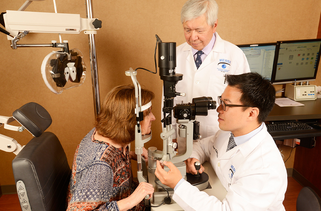
Symptoms alone rarely provide a definitive answer, which is why a comprehensive eye evaluation is so important.
At MERSI, a thorough evaluation for vision changes may include:
-
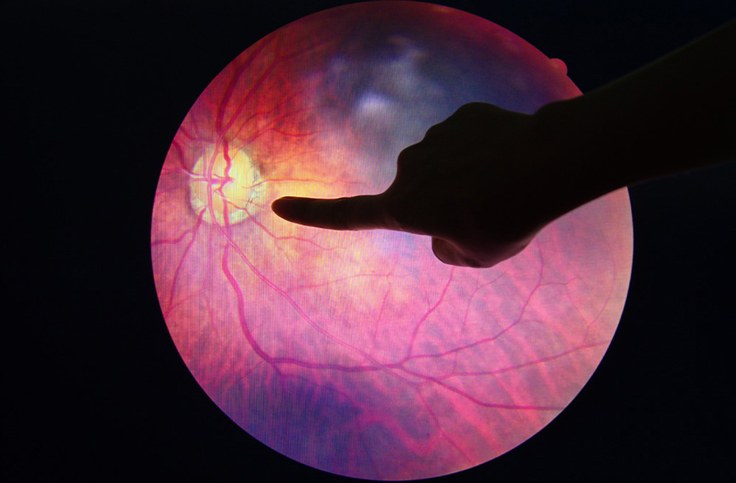
- Dilated eye exam: Widening the pupils allows physicians to examine the retina, macula, and optic nerve directly
- Optical Coherence Tomography (OCT): High-resolution imaging that captures cross-sectional layers of the retina, detecting microscopic fluid accumulation or swelling invisible to the naked eye
- Retinal imaging and fluorescein angiography: Dye-based photography that identifies leaking or blocked blood vessels
- Cataract assessment: Evaluation of lens opacity, density, and its impact on visual function
- Vision testing: Standardized measurements to assess the degree of functional vision loss
Self-diagnosing based on symptoms is genuinely difficult. The overlap between cataracts and retinal disease is significant enough that even experienced clinicians rely on diagnostic imaging rather than symptom patterns alone.
Schedule a Comprehensive Eye Evaluation at MERSI
Not all blurry vision is caused by cataracts, and not all retinal disease announces itself loudly. In many cases, the two conditions coexist, each influencing the other in ways that require expert evaluation to untangle.
A comprehensive eye evaluation at MERSI can help identify the true cause of your vision changes and guide a treatment plan designed for your specific situation. Our fellowship-trained specialists in Waltham, Massachusetts use advanced diagnostic imaging and a patient-centered approach to protect your vision at every stage.
If your vision has changed, gradually or suddenly, don’t wait to find out why. Schedule an evaluation at MERSI.
Frequently Asked Questions
Can retinal disease look like cataracts?
Yes. Retinal conditions such as macular degeneration and diabetic retinopathy can cause blurry or reduced vision that may initially resemble cataract symptoms. A comprehensive dilated eye exam and retinal imaging are needed to distinguish between the two.
Can cataracts hide retinal disease?
Yes. A dense cataract can obstruct a physician’s view of the retina, making it more difficult to identify underlying retinal conditions. This is one reason why a retinal evaluation is often recommended before cataract surgery.
Does diabetes cause blurry vision?
Yes. Diabetes can contribute to blurry vision in several ways, including blood sugar fluctuations, cataract development, diabetic retinopathy, and retinal swelling (macular edema). Patients with diabetes may mistake retinal symptoms for cataracts, which is why annual dilated eye exams are strongly recommended.
How do doctors determine whether blurry vision is caused by cataracts or the retina?
A comprehensive eye evaluation, including a dilated eye exam, OCT retinal imaging, and cataract assessment, can identify the underlying cause. Symptoms alone are often insufficient for an accurate diagnosis because the two conditions overlap considerably.
When should blurry vision be evaluated by an eye specialist?
Blurry vision should be evaluated promptly if it worsens over time, interferes with daily activities, occurs suddenly, or is accompanied by symptoms such as floaters, flashes of light, visual distortion, or dark spots. Sudden vision changes in particular warrant urgent care.